OUR SERVICES
MRI
An MRI is a diagnostic procedure that uses large magnets, radio frequency pulses and a computer to produce detailed images of organs and structures within the body.
CT Scan (CAT)
A computed tomography scan (CT or CAT scan), is a diagnostic imaging procedure that uses a combination of X-rays and computer technology to produce cross-sectional images (often called slices) of the body.
Ultrasound
An ultrasound, also called sonography, is a diagnostic imaging exam that uses a small transducer (probe) and ultrasound gel to expose the body to high-frequency sound waves to create images of blood vessels, tissues and organs.
X-ray
An X-ray is a diagnostic test that uses small doses of radiation to produce images of internal tissues, bones and organs onto film.
Arthrogram
Arthrography is the X-ray examination of a joint that uses fluoroscopy and a contrast material.
Bone Density
A bone density test, or DEXA scan, is a noninvasive procedure that uses X-rays to measure bone mass. The exam provides a measurement corresponding to the mineral density of bone, used to diagnose osteopenia and osteoporosis.
Fluoroscopy
Fluoroscopy is an imaging technique commonly used by physicians to obtain real-time images of the internal structures of a patient through the use of a fluoroscope.
Women's Services
Women's services are provided by board certified radiologists who sub-specialize in diagnostic mammography. These services are provided using state-of-the-art mammography equipment in an environment that is focused on women's health.
Mammography
Mammograms are one of the best ways to find the early stages of breast cancer. It can reveal small tumors up to two years before you or your doctor can feel them.
3D Mammography
Recommended for women with dense breasts or with a family history of breast cancer. Images are taken at the same time as a regular mammogram with the same system
Breast MRI
Magnetic Resonance Imaging (MRI) produces images using a strong magnet and radio waves. It is a sensitive test for the detection of breast cancer, but does not take the place of mammography.
Breast Ultrasound
Ultrasound imaging of the breast uses sound waves to produce pictures of the internal structures of the breast. It is primarily used to help diagnose breast lumps or other abnormalities your doctor may have found during a physical exam, mammogram or breast MRI.
GE 1.5T Signa HD
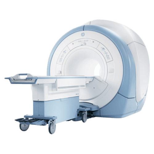
Downtown Columba
West Columbia
Weight limit 350lbs, bore width 54cm
Siemens 1.5T Espree Wide Bore
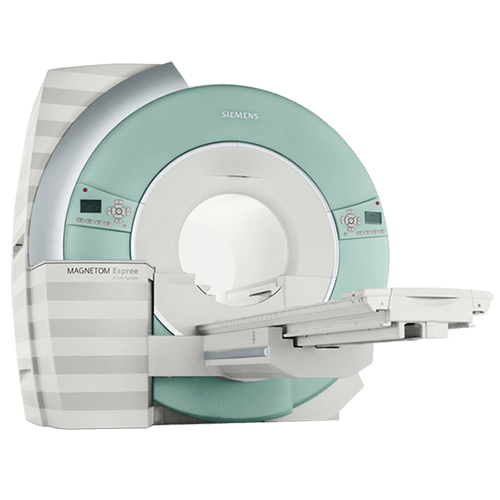
Downtown Columbia
Northeast Columbia at ImageCare
Weight limit 550lbs, bore width 70cm
Hitachi 1.2T Oasis Open MRI

Irmo
Weight limit 600lbs, gantry opening 45cm,
table top width 82cm
Physician Links
Patient Links
Get To Know Us
Interpretation services
Get To Know Us
Patient Links
Physician Links
Interpretation services
© Palmetto Imaging, Inc., All Rights Reserved
Translation services on this website are provided via Google™ Translate, a free automated translation service that can translate text into different languages. This tool is for your convenience only, and should not be considered exact and may in some cases include incorrect language. No warranty of any kind is made as to the accuracy, correctness, or reliability of any information translated by Google™ Translate. Please know that when a translation is requested, you will be leaving the the Palmetto Imaging website and any person or entity who relies on these translation services does so at his or her own risk. If you have any questions about Google™ Translate, please click the following link: Google™ Translate FAQs.